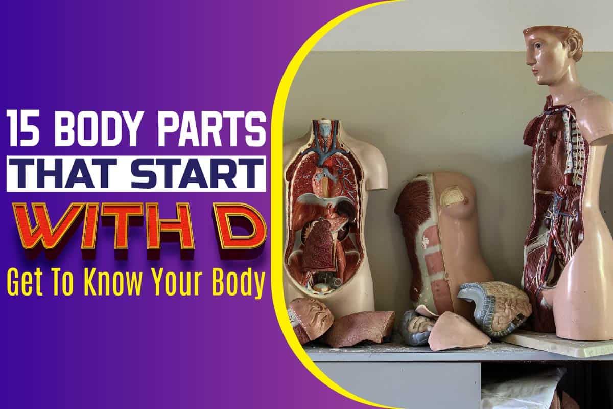
If you were asked to name the body part that starts with the letter “d”, how many would you recite by heart?
You’ll probably not be able to recite many because some might be strange to you. After all, they are inside of you and out of sight.
If you were searching for the body part that starts with the letter “d”, we’ve prepared a list for you explaining what they do.
Diaphragm
This body part is a thin skeletal muscle that sits between the chest and the stomach. It is linked to the spine and surrounded by the ribs and sternum.
The diaphragm helps you to breathe properly. As you inhale, your diaphragm flattens and contracts.
Your rib cage will begin to rise when this happens; likewise, your lungs will be filled with air. However, once you exhale, your ribs will lower, and your diaphragm will relax.
Descending Colon
The colon is a part of the large intestine and the digestive system. It helps to process waste products and prepares them for removal, and also reabsorb fluids.
The descending colon has four parts, namely;
- ascending colon
- descending colon
- sigmoid colon
- transverse colon
Furthermore, while the small intestine absorbs nutrients from your food, the large intestine doesn’t have much to do. It helps to maintain water balance, stores waste and absorbs specific vitamins.
You can find the descending colon on the left part of the large intestine. It extends to the sigmoid colon. Peritoneum, a thin tissue layer supporting the abdominal organ, is what holds the colon in place.
Lymph vessels, nerves and blood vessels all go through the peritoneum to reach several organs.
Duodenum
This is a part of the small intestine that takes the shape of the letter “C”. Furthermore, it is close to the lumbar section and the kidney. It’s the first section of the small intestine that is linked to the jejunum.
Once you’ve eaten food, the food travels to your stomach before going through the Duodenum.
The Duodenum plays a major role in breaking down the partially digested food from the stomach and extracting nutrients from them.
After it has done its job, the food now finds its way to the small intestine.
Dermis
The dermis is the layer of your skin that sits underneath the outer layer called the epidermis. The role of the dermis is to provide nutrition to the epidermis because the outer layer is not equipped to provide food for itself.
Furthermore, the dermis comprises all the sweat glands, blood capillaries, hair follicles, and nerve endings. It is hearty, thick and protects your internal organs.
Also, the sweat glands present in the dermis control sweat production in response to specific conditions like stress and heat and anxiety.
As the sweat gets off the skin, the body begins to cool down and maintain homeostasis.
Digits
Your toes and fingers are known as digits. The bones in the digits are known as phalanges, and the origin of digits makes us understand that it isn’t all animals that possess this body part.
Digestive system
The digestive system is specially designed to transform the food that we eat into nutrients. Furthermore, the body uses these nutrients for cell repair, energy and growth.
Here’s how it works
Mouth
The activity of the digestive system begins with the mouth. As soon as you take in food, chew and swallow, the process has begun.
The food gets broken down into smaller particles that can easily digest. Saliva mixes with the food to start the breaking down process into an acceptable form that your body can use.
Throat
Once food goes through your mouth, its next destination is the throat. From the throat, it moves further to the swallowing tube or oesophagus.
Oesophagus
This is a tube that extends from the throat to the stomach. In essence, the oesophagus transports food to the stomach.
Stomach
The stomach is an organ with solid muscular walls. Apart from storing food, it serves as a grinder and mixer. The stomach discharges acid and potent enzymes that continuously breaks down food.
Once food leaves the stomach, it is either in liquid or paste form. From there, food transports to the small intestine.
Small intestine
The small intestine continues from where the stomach stopped. It breaks down food by leveraging the bile in the liver and the enzymes in the pancreas.
From the small intestine, it gets to the pancreas, liver, gall bladder, colon, rectum and anus.
Ducts
Ducts are the part of the body where tears come from. They are little tunnels that are designed to transport tears. Ducts are linked to your throat.
Furthermore, ducts help drain excess liquid within the area whenever you get emotional and cry; the ducts transport water (tears) through your eyelids.
Dua’s Layer
This is one of the six layers that you can find in the cornea. Remarkably, the Dua’s layer body part was just discovered in 2013. It is a very thin layer.
Harminder S. Dua, a professor of visual sciences and ophthalmology, discovered the Dua’s layer.
The Dua’s layer is a well-defined, tough lining about 10 to 15 micrometre thick, located between the Descemet’s membrane and the corneal stroma.
The layer helps surgeons to improve the results of patients who undergo transplants and corneal grafts.
During surgery, little air bubbles are introduced into the corneal stroma. This is the “big bubble technique”.
In most cases, the bubble can erupt and damage the patient’s eye. However, if the surgeon injects the air bubble under the Dua’s layer and not above it, the layer’s strength will reduce the possibility of tearing.
Dorsal Cavity
This is a section of the body that is filled with fluid. It surrounds the spinal cord and the brain.
It comprises the spinal cavity and cranial cavity, both of which offer protection to the delicate nervous tissue.
The spinal cord is a fragile body part that needs the protection of the Dorsal cavity.
Dorsalis Pedis Artery
The dorsal pedis, also known as the artery of the foot, is the blood vessel found in the lower limb. It transports oxygenated blood to the tip of the foot.
In most cases, it is examined by doctors to check if a patient is suffering from peripheral vascular disease.
Derriere
Derriere is a French word for buttocks or backside. The Derriere refers to the two rounded portions of our lower backside.
Humans use it for sitting on chairs.
Dura Mater
This is one of the layers that assist in covering the brain and spinal cord. It’s the topmost layer among the three meninges that offer protection to the brain and spinal cord.
Furthermore, the Dura Mater supports and surrounds the large dural sinuses (venous channels), transporting blood from the brain to the heart.
The Dura Mater is divided into numerous septa that supports the brain. Also, it is thick and uses connective tissues to produce the largest possible protective layer for the nervous system beneath it.
Decidua
The Decidua is a protective uterine tissue that plays an essential role in shielding the embryos from getting attacked by motherly immune cells.
It also offers nutritional support to the emerging embryo before the formation of the placenta.
The major functions of the Decidua are the regulation of syncytiotrophoblast invasion, provision of gas exchange and nutrition and the production of hormones.
To prepare the uterus for pregnancy, the decidualization process must commence.
This is the preparation of the innermost layer of the endometrium by using spiral arterioles and trophoblast to invade it.
The Decidua is separated into three parts. The decidua capsularis, decidua basalis and the decidua parietals.
These three parts are named by the type of relationship that they have with the conceptus.
Descending Aorta
The aorta is the largest artery in the body. It runs through the abdomen and the chest. The descending aorta helps transport blood to various parts of your body.
Furthermore, the aorta is the primary artery that moves blood from your heart to other parts of the body. The blood is flown out of the heart via the aortic valve.
Moving on, the blood moves through the aorta in a cane-shaped bend that enables other primary arteries to distribute oxygen-rich blood to the muscles, brain and other cells for the body to survive.
Deltoid
The deltoid muscle is responsible for creating the rounded contour that is visible on the human shoulder.
Another name for the deltoid muscle is the common shoulder muscle, mostly seen in domestic cats’ anatomy.
The deltoid muscle consists of three different types of muscle fibres, and they are as follows;
- clavicular or anterior part
- scapular or posterior part
- acromial or intermediate part
Diencephalon
This is a little part of the brain that is hidden from view when you’re staring at the outer part of the brain. It’s divided into four sections, namely;
- thalamus
- epithalamus
- hypothalamus
- subthalamus
- Diaphram
The diaphragm is a muscular structure that divides the abdominal cavities and chest (thoracic) in mammals. It is the primary respiration muscle.
Similar Posts:
- Why Am I Hungry After I Eat?
- Why Does My Stomach Hurt Every Morning?
- Why Do My Hands Swell When I Walk?
- Why Do Beans Make You Fart?
- Alcohol’s Impact on the Body and Mind
- Why Do You Feel Better After You Throw Up?
- Why Do I Get Dizzy When I Bend Over?
- Why Does Coffee Make Me Sleepy?
- Why Is My Thumb Twitching?
- Why Do I Yawn When I Workout?

